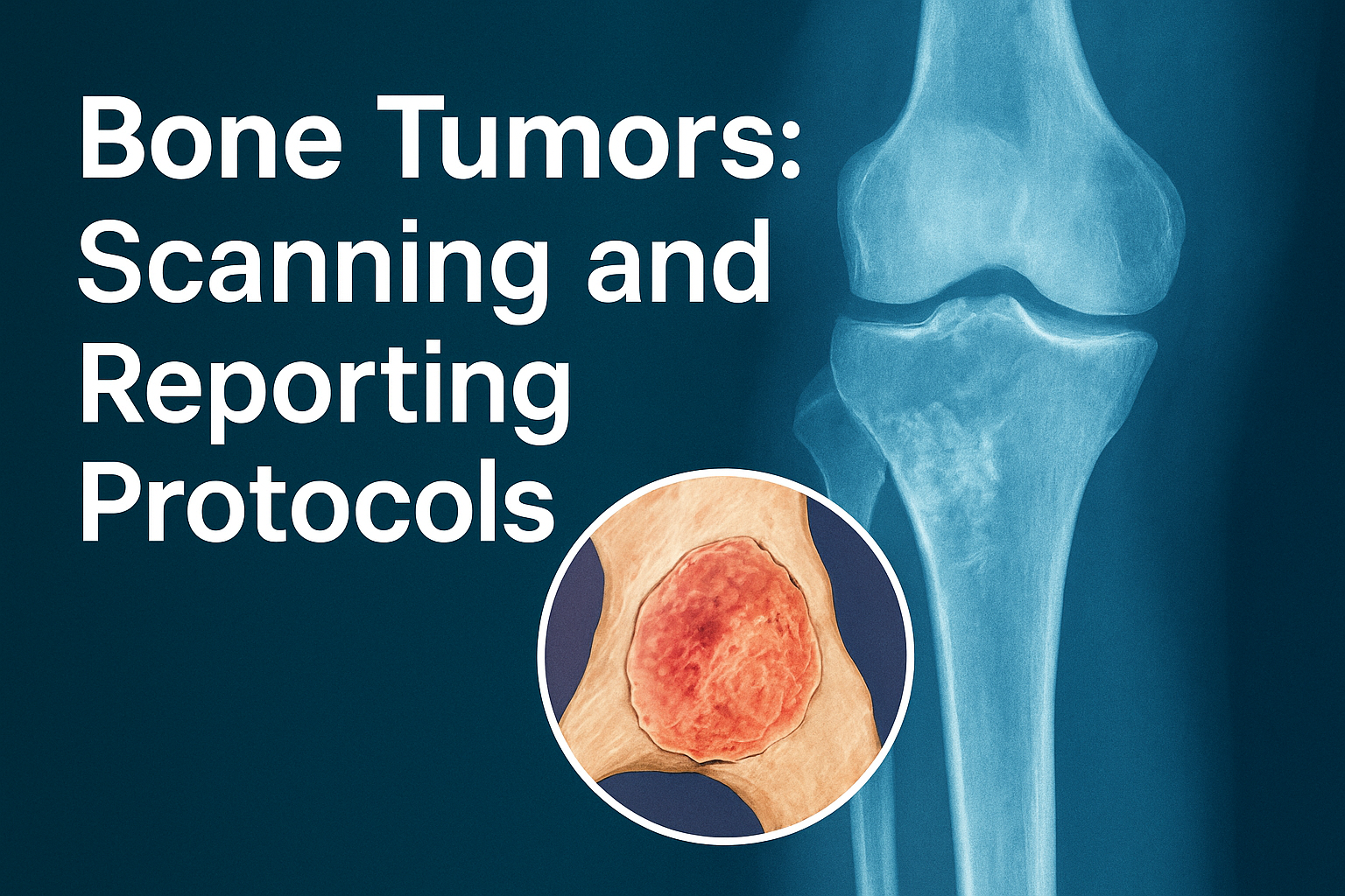
Bone tumors are one of the most challenging conditions in musculoskeletal (MSK) radiology. For both patients and doctors, accurate diagnosis is the foundation of effective treatment. The role of a radiologist is not just to detect a lesion but also to differentiate between benign and malignant tumors, characterize the lesion precisely, and guide management.
At Izen Imaging & Interventions, Noida, we specialize in advanced MRI, CT, PET/CT, and X-ray imaging protocols for bone tumors. Our goal is to ensure nothing critical is missed, providing clarity and confidence in every report.
Why Imaging Protocols Matter in Bone Tumors
Imaging plays a pivotal role in the diagnosis and treatment pathway of bone tumors. A structured approach offers multiple benefits:
- Accurate Staging: Imaging helps determine the size, spread, skip lesions, joint involvement, and neurovascular encasement.
- Guiding Biopsy: Proper imaging protocols highlight where and how to biopsy, preventing sampling errors.
- Monitoring Treatment: MRI is vital for assessing post-chemotherapy necrosis and evaluating treatment response.
- Avoiding Misdiagnosis: Certain conditions mimic tumors (such as infection or benign “leave-alone lesions” like enchondroma or non-ossifying fibroma). Imaging prevents unnecessary surgeries.
Step 1: The Role of Radiographs (X-rays)
The golden rule in bone tumor imaging: Never interpret an MRI without an X-ray.
- Zone of Transition: A narrow zone usually means a benign lesion, while a wide zone suggests aggressiveness.
- Periosteal Reaction: Thick, smooth reactions are benign, whereas irregular or sunburst patterns point to malignancy.
X-rays form the foundation for deciding the next steps in tumor characterization.
Step 2: MRI – The Workhorse of Bone Tumor Imaging
When is MRI Needed?
MRI is the most comprehensive tool for bone tumor evaluation. It is used for:
- Staging and preoperative planning
- Cases where radiographs are equivocal
- Differentiating benign from malignant lesions
- Screening for skip lesions
Our MRI Protocol at Izen Imaging
We follow an internationally benchmarked MRI protocol:
- Coronal STIR and T1 for marrow evaluation
- Sagittal T2 for structural detail
- Axial STIR, T1, and T2 for lesion characterization
- Dynamic contrast-enhanced MRI (DCE-MRI) with mean curve analysis
- Post-contrast T1 fat-sat in three planes
- Large FOV T1 covering the joint above and below the lesion, along with whole-limb screening
This combination ensures that both local detail and systemic staging are addressed, leaving no room for missed skip lesions or subtle spread.
Step 3: Structured Reporting – What Should Be Included?
A standardized bone tumor report improves decision-making for surgeons and oncologists. It should include:
- Signal characteristics on T1, T2, and STIR
- Enhancement patterns and percentage of necrosis
- Lesion dimensions – maximum length and transverse diameter
- Relation to joints and landmarks
- Neurovascular bundle involvement
- Transphyseal or intra-articular extension
- Presence of skip lesions
This structured approach ensures clarity for the referring clinician and avoids ambiguity.
Step 4: Pitfalls and Common Misses
Bone tumor imaging has its challenges. A few key pitfalls include:
- Mimics: Infections, stress fractures, and Langerhans cell histiocytosis can look like tumors.
- Ignoring marrow edema: If unexplained, further CT or PET/CT is warranted.
- Older patients (>35 years): Always consider metastasis, myeloma, or lymphoma before labeling a lesion as primary bone tumor.
- Over-interpreting incidental lesions: Some benign lesions, like enchondroma or fibrous dysplasia, require no treatment (“leave-alone lesions”).
Step 5: Advanced Imaging – Dynamic Contrast MRI
One of the most powerful tools in bone tumor imaging is Dynamic Contrast-Enhanced MRI (DCE-MRI).
- Benign tumors usually show Type 1 or 2 curves (slow or progressive enhancement).
- Malignant tumors typically show Type 3 or 4 curves (rapid uptake with plateau or washout).
- Osteoid osteoma, for example, demonstrates a characteristic arterial enhancing nidus, which is highly specific.
DCE-MRI adds only 4–5 minutes to scan time but provides invaluable physiological as well as morphological information.
Minimally Invasive Biopsies & Interventions at Izen Imaging
At Izen Imaging & Interventions, Noida, our role goes beyond just diagnosis. We specialize in a wide range of image-guided procedures that make patient care smoother, faster, and more precise.
- CT-guided biopsies – Essential for sampling deep-seated bone and soft tissue lesions with pinpoint accuracy.
- Ultrasound-guided biopsies – A less invasive, real-time technique for superficial and accessible lesions.
- Therapeutic procedures – Including aspiration of cystic lesions, drainage procedures, and image-guided injections for pain relief or local tumor control.
What sets us apart is the way these procedures are performed:
- All interventions are done under safe anesthesia coverage, ensuring patient comfort.
- They are carried out on an OPD (outpatient) basis, reducing hospital stay and overall costs.
- With our expertise, procedures are minimally invasive, less expensive, and recovery is quick.
Importantly, we are active members of multiple bone tumor committees, which means we are regularly updated on the latest guidelines and best practices. This allows us to perform biopsies in a way that maximizes diagnostic yield, minimizes complications, and directly benefits the patient’s treatment plan.
By combining advanced imaging with expert interventional radiology, Izen Imaging ensures that patients not only get the right diagnosis but also safe, affordable, and effective procedures under one roof.
Key Takeaways for Patients and Doctors
- Always begin with X-rays before MRI.
- MRI provides unmatched detail for staging, skip lesions, and neurovascular mapping.
- A structured reporting system ensures clear communication.
- DCE-MRI should be included whenever contrast is given for bone tumors.
- Recognize benign “leave-alone lesions” to avoid overtreatment.
- In patients over 35, metastasis or myeloma must be considered first.
Conclusion
Bone tumor imaging is not just about identifying a lesion — it is about guiding the entire clinical journey from diagnosis to treatment. At Izen Imaging & Interventions, Noida, we combine advanced MRI protocols, structured reporting, and minimally invasive biopsy options to provide comprehensive bone tumor care.
If you or your loved one has been advised a bone tumor scan, choose a center that offers technology with expertise and compassion.
Visit us at Izen Imaging & Interventions, Noida, for advanced bone tumor imaging and reporting you can trust.
Written by – Dr. Khushboo Pilania
Posted by – Falak Ali