
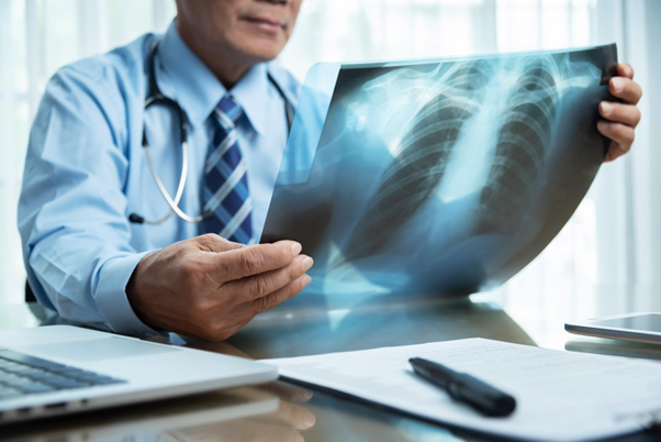
What is a Chest CT Scan?
A chest CT scan, also called thoracic CT or computed tomography of the chest, is an advanced diagnostic imaging test that creates detailed cross-sectional images of the chest area. It utilizes X-rays and sophisticated computer technology to generate highly detailed images of the chest, encompassing the lungs, heart, blood vessels, airways, oesophagus, and other vital structures. It produces multiple cross-sectional slices, delivering clearer and more comprehensive information than traditional X-rays. This capability assists healthcare providers in diagnosing and assessing various chest-related conditions.
Uses of Chest CT Scans
Chest CT scans are crucial for diagnosing and evaluating a wide array of conditions, including:
- Lung Conditions: Such as pneumonia, lung cancer, pulmonary embolism, and interstitial lung diseases.
- Cardiovascular Conditions: Including heart disease, coronary artery disease, and aortic aneurysms.
- Chest Trauma: Assessing injuries to the chest wall, lungs, or other structures following trauma or accidents.
- Infectious Diseases: Evaluating infections like tuberculosis or fungal infections affecting the lungs.
- Structural Abnormalities: Detecting anomalies in the chest wall, airways, or blood vessels.
- Monitoring Treatments: Tracking the effectiveness of treatments for lung or heart conditions over time.
Procedure for a Chest CT Scan
- Preparation: Depending on the type of scan, you may need to change into a hospital gown and remove metal objects that could interfere with imaging. Fasting for a few hours might also be required.
- During the Scan: You'll lie on a motorized table that slides into the CT scanner, a large, doughnut-shaped machine. Staying still during the scan is crucial for clear images, despite the buzzing or clicking noises emitted by the scanner.
- Contrast Injection: In some cases, a contrast dye (iodine-based) may be injected into a vein in your arm to enhance visibility of blood vessels and other structures, improving image clarity.
- Post-Scan: After the scan, you can usually resume normal activities unless advised otherwise by your doctor. The images are interpreted by a radiologist, who provides a report to your healthcare provider.
Benefits of Chest CT Scans
- Accuracy: Provides detailed images that can reveal abnormalities not visible on conventional X-rays.
- Speed: The procedure is usually quick and painless, lasting only a few minutes.
- Guidance for Treatment: Helps healthcare providers make informed decisions regarding treatment by accurately diagnosing conditions and monitoring their progression.
Chest CT scans are invaluable in diagnosing and monitoring various chest conditions, offering detailed insights crucial for treatment planning. Despite minimal risks, the benefits of precise diagnosis and monitoring often outweigh concerns. Always discuss any questions or concerns with your healthcare team for personalized guidance.